
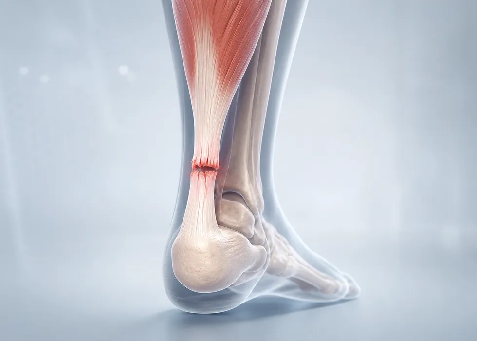
The Achilles tendon is the thick cord that connects the calf muscles to the heel. It is essential for walking, climbing stairs, jumping, and sudden acceleration. A rupture means the tendon fibers have torn partially or completely, most often during an abrupt push-off movement in sports such as football, basketball, tennis, or recreational running.
Who Is Most at Risk?
Achilles tendon rupture is especially common in adults between 30 and 50 years old who are not training consistently but perform intense activity intermittently. This is often described as the “weekend warrior” pattern. Risk increases further when one or more of the following factors are present:
- Previous Achilles pain or chronic tendon degeneration
- Fluoroquinolone antibiotic use
- Corticosteroid injections around the tendon
- Diabetes, inflammatory disease, or metabolic disorders
- Reduced calf flexibility and poor conditioning
One reason the tendon is vulnerable is that the segment roughly 2 to 6 centimeters above the heel has relatively limited blood supply. When repeated microtrauma accumulates in that area, the tissue becomes less capable of repair.
Typical Symptoms
Many patients describe a sudden snap or pop, followed by the sensation that someone kicked the back of the ankle. Common findings include:
- Sharp pain at the back of the ankle or lower calf
- Swelling and bruising
- Weak push-off while walking
- Difficulty standing on tiptoe
- A visible or palpable gap in some cases
Because some smaller muscles can still move the ankle, Achilles rupture is sometimes mistaken for a severe ankle sprain. That is why examination by an orthopedic specialist is important when the story is suspicious.
How Diagnosis Is Made
Diagnosis begins with the history of injury and a focused physical examination. One of the best-known clinical tests is the Thompson test: when the calf is squeezed, the foot should point downward. If that movement is absent or clearly weakened, a rupture is strongly suspected.
Ultrasound or MRI may be requested when the diagnosis is unclear, when there is concern for a complex tear, or when surgical planning requires more detail. Imaging is also helpful in chronic or delayed presentations.
Modern Treatment Approach
Management is no longer a simple “surgery versus no surgery” debate. The most important insight from modern studies is that structured functional rehabilitation changes outcomes significantly.
Non-operative treatment
For many patients, especially those with lower athletic demands or higher surgical risk, treatment can be successful without surgery. The tendon is protected in a boot with heel elevation, and weight-bearing plus motion are advanced gradually under a supervised rehabilitation plan.
Benefits include avoiding wound problems, infection, and anesthesia-related risk. Non-operative care can be an excellent option when the tendon ends are well aligned and the patient can follow the rehab schedule closely.
Surgical treatment
Surgery may be preferred in selected younger, highly active, or professional athletes, as well as in cases with large tendon separation or delayed presentation. The tendon ends are repaired and then protected while healing progresses. Different techniques exist, including open and minimally invasive repair.
The potential advantage is a strong anatomical repair, but surgery also carries risks such as wound healing problems, infection, or nerve irritation.
Rehabilitation Matters as Much as the Initial Treatment
Regardless of whether the rupture is treated surgically or non-operatively, recovery depends heavily on rehabilitation. The early goal is to protect the repair while avoiding unnecessary stiffness and calf atrophy. Later phases focus on:
- Restoring ankle motion
- Rebuilding calf strength
- Recovering balance and proprioception
- Progressing to sport-specific loading
Return to full athletic activity commonly takes several months, and high-level explosive sports may require closer to 6 to 12 months.
When to Seek Medical Attention
Urgent evaluation is appropriate if you feel a pop in the tendon area, cannot push off normally, or suddenly lose the ability to rise onto your toes. Early diagnosis improves treatment planning and reduces the risk of prolonged weakness or chronic tendon dysfunction.